
We use the latest technology to help you see better!
Optomap®
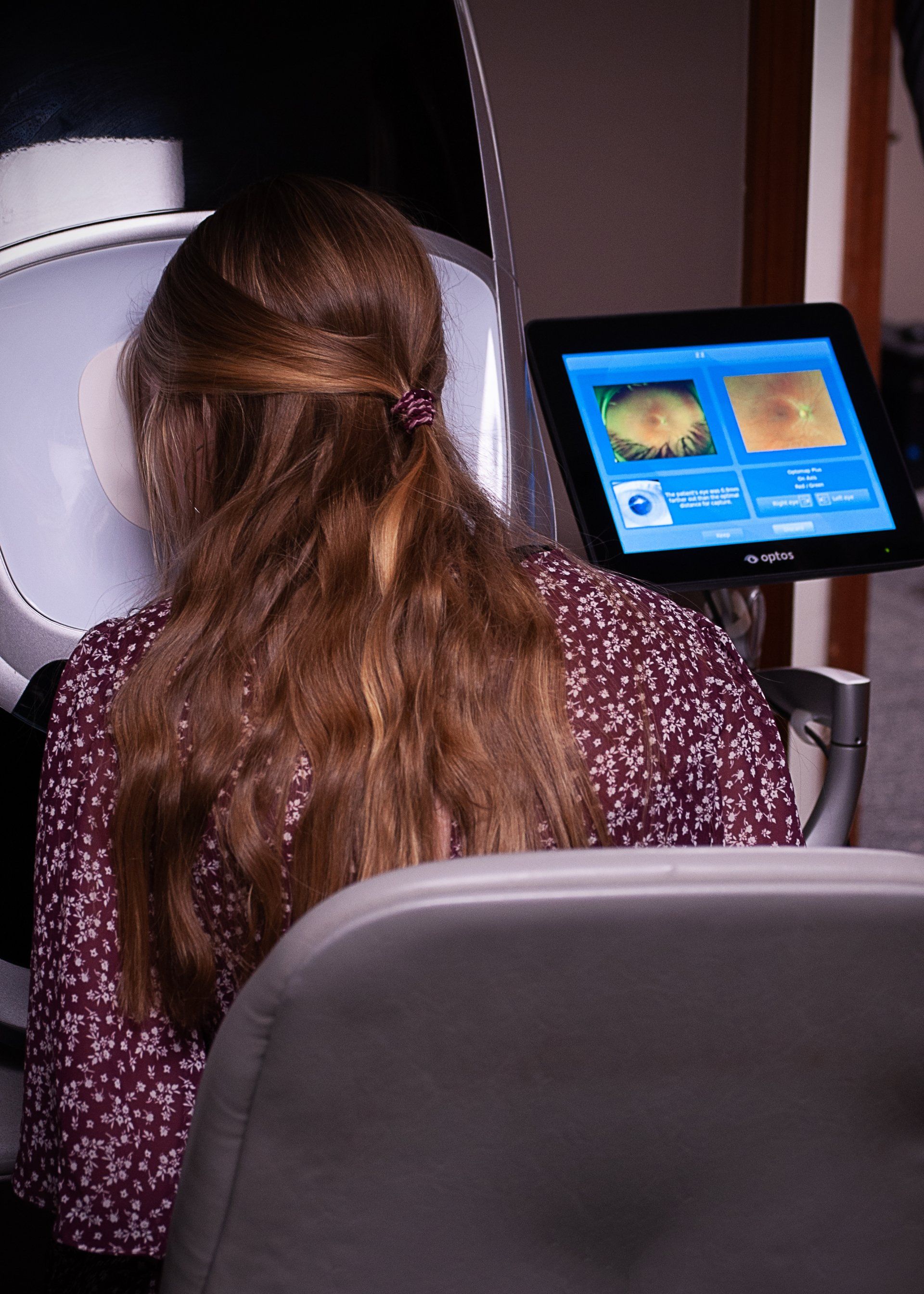
Annual eye exams are vital to maintaining your vision and overall health. Atchison Eye Center offers the optomap® as an important part of our eye exams. The optomap produces an image that is unique and provides Dr. Franken with a high-resolution 200° image in order to ascertain the health of your retina. This is much wider than a traditional 45° image. Many eye problems can develop without you knowing, in fact, you may not even notice any change in your sight – fortunately, diseases or damage such as macular degeneration, glaucoma, retinal tears or detachments, and other health problems such as diabetes and high blood pressure can be seen with a thorough exam of the retina.
The inclusion of optomap as part of a comprehensive eye exam provides:
A scan to show a healthy eye or detect disease.
A view of the retina, giving your doctor a more comprehensive view than they can get by other means.
The opportunity for you to view and discuss the optomap image of your eye with your doctor at the time of your exam.
A permanent record for your file, which allows Dr. Franken to view your images each year to look for changes.
The optomap is fast, easy, and comfortable for anyone. The entire image process consists of you looking into the device one eye at a time. The optomap images are shown immediately on a computer screen so we can review it with you.
Schedule your optomap today!
For more information on the optomap please visit the optomap website.
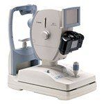
TRS 5100
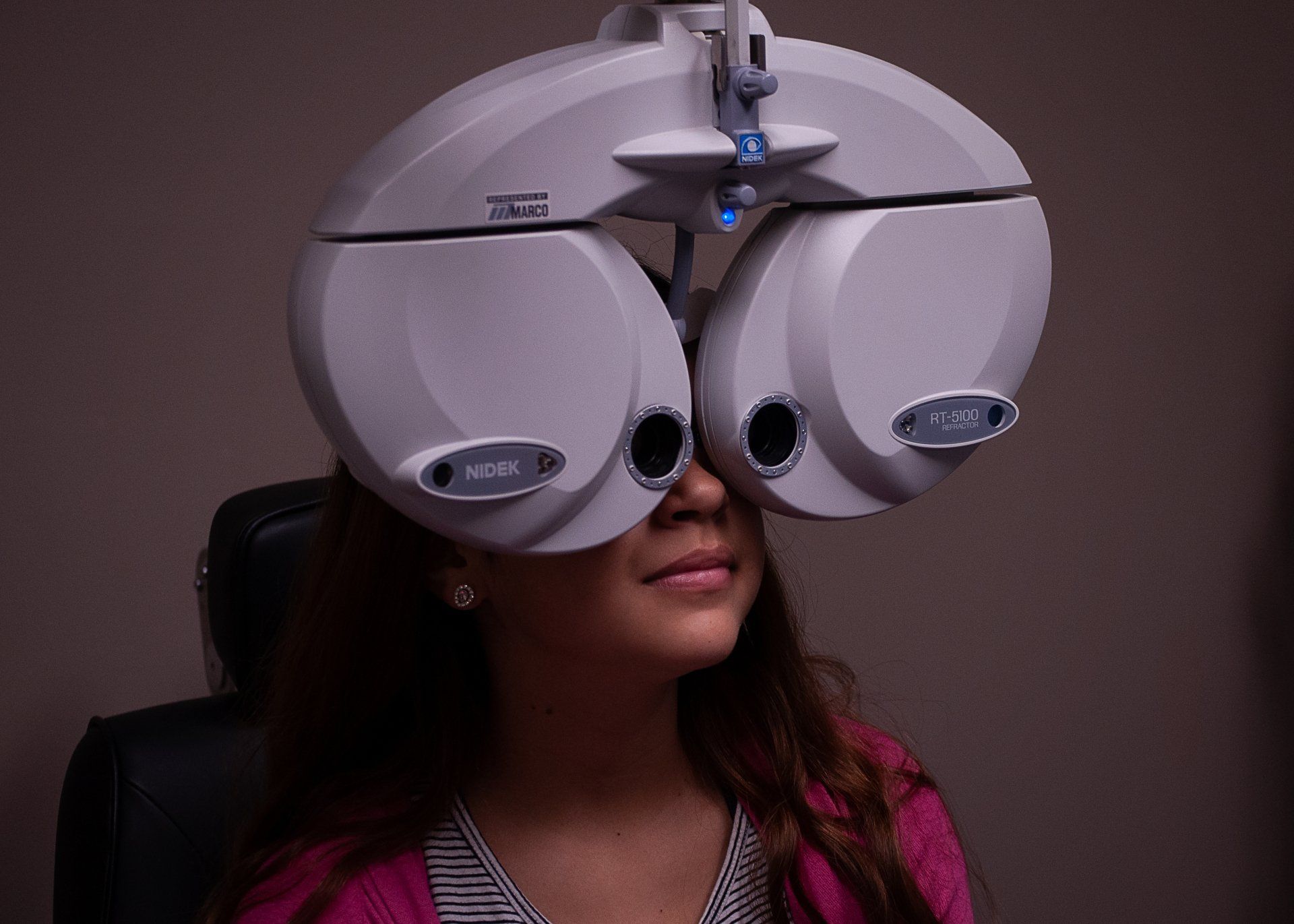
Our office is committed to offering each of our patients the finest in eye care. You’ll see how your overall experience will be enhanced comparing your old vs. new Rx with a push of a button and no more answering the question, “Do you like 1 or 2 better?” Our state-of-the-art automatic refractor allows us to achieve the most accurate prescriptions to help you see your best in a fraction of the time.
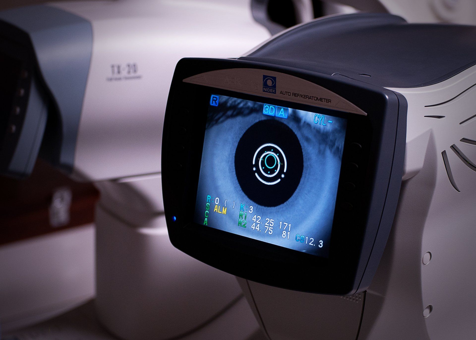
Autorefractors are machines that automatically determine the correct lens prescription for your eyes. If you’ve discovered you might need vision correction during your eye examination, it’s vital to determine just how “much” your eyes need to be corrected with lenses or contact lenses. This is called measuring your “refraction.” Autorefractors automatically measure this value during an eye examination.
Autorefractor/
Autokeratometer
Digital Fundus Photography consists of a digital camera system that takes a photograph of your retina. Diseases such as macular degeneration, glaucoma, retinal tears or detachments, as well as other health problems such as diabetes and high blood pressure, can be detected with a thorough exam of the retina.
Non-Mydriatic Retinal Camera
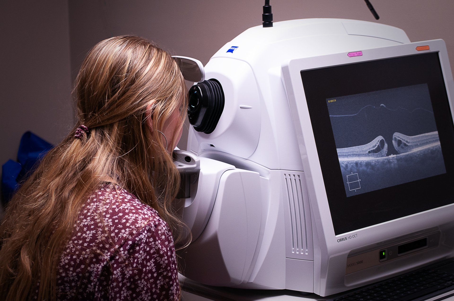
Optical Coherence Tomography (OCT) is a non-contact medical imaging technology similar to ultrasound and MRI. With OCT, reflected light is used to produce detailed cross-sectional and 3D images of the eye.
Spectral Domain Technology
The visual field is a diagnostic device used to test a patient’s complete “visual field,” which includes the peripheral vision. It is a simple, painless, out-patient procedure and takes only minutes. This test is invaluable in diagnosing, and monitoring the treating patients with glaucoma, where peripheral vision irreversibly lost without symptoms until later stages.
Visual Field Analyzer
Non-contact (or air-puff) tonometry does not touch your eye but uses a puff of air to flatten your cornea. It is often used as a simple way to check for high IOP and is the easiest way to test children. It may also be used for people who have had LASIK surgery. Noncontact tonometry does not use numbing eyedrops.